EXHIBITOR SHOWCASES
FEI Company
Friday, July 22, 2016, 12:00 – 13:00, Forum Hall
3D Isotropic Volume Imaging and Reconstruction of Biological Samples – Enhancing Resolution in X, Y and Z
Wim Voorhout (Netherlands)
Visualizing the three-dimensional (3D) architecture of cells and tissues is essential for understanding relationships between structure and function in biological systems. FEI Teneo VolumeScope is a novel Serial Block Face Imaging (SBFI) solution for high spatial resolution and throughput SEM volume imaging overcoming the resolution limits set by mechanical slicing by combining it with optical sectioning, realized by Multi-Energy Deconvolution SEM (MED-SEM). Teneo VS tightly integrates SBFI and Multi-Energy Acquisition while automating and streamlining the complete 3D imaging workflow. The system can analyze large area/volume acquisitions by automatically tiling multiple fields of view into a large composite image of each block. It can also import and overlay images from light microscopes to allow direct targeting of regions of particular interest based on fluorescence staining or other LM techniques. An automated, in-chamber microtome can be easily removed to permit for other SEM applications. A newly designed in-column detection system provides high-efficiency, high-contrast images of biological materials. Teneo VS can be operated in high-vacuum mode to achieve the highest resolution or low-vacuum mode with water vapor in the chamber to mitigate sample charging efficiently. The final results can be instantly and easily visualized using Amira Live Tracker for on the fly analysis and subsequently analyzed using the world’s leading 3D imaging software, Amira.
Amira for Cell Biology – Flexible Segmentation and Object Tracking with Amira
Jan Biesebrecht (Germany)
One of the main challenges for today’s cell biologist is efficient and flexible processing of time series data to follow and study any type of intra- and inter-cellular processes. With Amira for Cell Biology, we are enabling these researchers to apply custom-workflows designed to segment their data efficiently. Our collaboration with the Danuser laboratory enables Amira to apply the most accurate object tracking solution available to researchers, directly to the segmented data to obtain precise information about the processes of interest. In this vendor tutorial, we will be demonstrating how to apply Amira’s flexibility in creating custom workflows to different types of data that can then be tracked with Amira’s object tracking solution powered by U-Track.
Leica Microsystems
Saturday, July 23, 2016, 12:00 – 13:00, Forum Hall
Leica SP8 HyVolution Turns Super-sensitivity into 140 nm Resolution
Daniel Smeets (Germany)
Key Words: super-resolution, sensitivity, DNA nanorulers, live cell imaging.
In this presentation, we demonstrate how we improve the resolution of the Leica TCS SP8 confocal microscope using super-sensitive HyD hybrid detectors and Huygens deconvolution in our new HyVolution system. Compared to the usual diffraction limit around 240 nm, we achieve sub-diffraction limited resolution of 140 nm laterally and an axial resolution increase by a factor of 2.
This is possible, because of the high signal-to-noise ratio of the HyD detector. Being a photon-counting detector, Leica HyD circumvents noise amplification associated with traditional intensity averaging. Its superior signal-to-noise ratio compared to (GaAsP) photomultipliers thus translate into higher resolution when combined with state of the art deconvolution. The main advantage of using HyVolution over other techniques addressing this resolution domain is that you can freely choose to acquire multiple colors simultaneously at full speed. This makes it ideal for fixed specimen and live cell imaging alike.
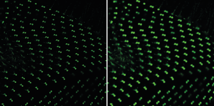
Underline: Basal bodies of Paramecium ciliae. HyVolution reveals ring-like structure. Sample courtesy by Anne Aubusson-Fleury, CNRS Gif-sur-Yvette, France.
Cell and Tissue Imaging by Confocal Microscopy, Sted and SHG Techniques in Cancer Research
Lucie Kubinova (Czech Republic) and Oleksandr Chernyavskiy (Czech Republic)
Key Words: confocal microscopy, super-resolution, second harmonic generation.
In this presentation, we demonstrate and compare a number of microscopic techniques for cell and tissue imaging using Leica confocal microscope systems including Leica SP8 with multiphoton excitation and Leica SP8 STED.
First, we will present images of microtubules (MTs) of human osteosarcoma cells acquired by super-resolution as well as confocal microscopy. We will compare resolution of MTs obtained by different microscopic techniques and image processing algorithms improving resolution, such as deconvolution.
Further, we will show imaging of unstained melanoma and colon tumor tissue samples by confocal one-photon and two-photon microscopy including analysis of collagen morphology visualized by second harmonic generation (SHG) and laser scanning confocal microscopy in reflectance mode. It will be demonstrated that such imaging techniques present a powerful tool for non-invasive in vivo studies.
This study was supported by MEYS (LM2015062 Czech-BioImaging).
Nanolive
Sunday, July 24, 2016, 12:00 – 13:00, Forum Hall
The 3D Cell Explorer: A revolutionary Tomographic Microscope to look instantly inside living cells in 3D!
Luca Clario (Switzerland)
Every Cell is unique and has its own complex structure.
We have developed a disruptive technology which, for the first time, allows users to explore instantly the inside of a living cell in 3D without the need for any labeling or any other invasive methods.
The 3D Cell Explorer is a high speed, high resolution and non-invasive tool that can look deep inside biological systems. This allows you to record stunning 3D images of entire cells in just seconds and with a higher resolution than any conventional microscope on the market.
With the 3D Cell Explorer, researchers, students and medical doctors can directly experience what happens inside the living cell – in real time!
NEWS
July 24
Saturday photos has been posted..
July 19
Practical information fully updated.
July 19
A - Z information available.
July 19
Check out the content of our Exhibitor Showcases.
July 15
Details on Congress Dinner published. Tickets will be available at registration desk.
July 1
Just announced! Symposium “Cell Biology Education – Teaching and Training in Research and Scientific Writing”
June 28
Best Poster Award!
June 22
Detailed Scientific programme and Guidelines for authors published.
SPONSORS
![]()
![]()
![]()

EMBO
excellence in life sciences
![]()
CONGRESS ORGANISED BY
![]()
to the
Congress Start
Czech Society for Cell Biology, z.s., Albertov 4, 12800 Prague, Czech Republic
Home | Sitemap | http://www.cscb.cz | info@iccb2016.org
Copyright © 2014 iccb2016.org. Powered and created by E-WORKS - web studio | XHTML 1.0 | CSS 2